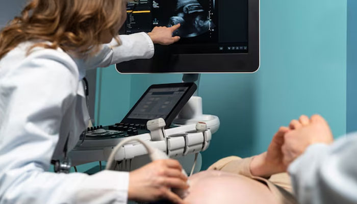
Experts highlight uses of ultrasound in diagnosis, treatment of diseases
Ultrasounds are one of the most useful tools in caring for women during pregnancy, childbirth and the post-delivery recovery period. The procedure allows gynaecologists to properly assess early stages of pregnancy, fetal growth and fetal well-being, providing doctors with diagnostic abilities that can greatly benefit obstetric patients.
These view were shared by Prof Sonia Naqvi, fellow of the Royal College of Obstetricians and Gynaecologists and fertility expert, at a recently held public health awareness seminar titled ‘Ultrasound: A Miracle in Medicine’.
Sje said the well-being scan, which is the most widely recommended scan, is offered for a number of reasons. It determines the lie or presentation of the baby (breech, transverse or cephalic), provides information about the placenta location and appearance, establishes an estimated fetal weight, examines the umbilical cord Doppler flow to determine whether the placenta is functioning as it should, calculates the amniotic fluid volume around the baby and allows for the sonologists to check the standard fetal measurements.
Time is also taken to make sure that the baby is swallowing, breathing and moving normally, explained by Prof Sonia. Earlier, Dr Nadia Farhan Essa, consultant family physician and chief operating officer at the Dr Essa Laboratory & Diagnostic Centre, inaugurated the seminar and appreciated the healthcare professionals including local obstetricians, radiologists, general practitioners, sonologists and other allied health professionals.
Highlighting the recent studies in diagnostic sonography, she said that according to the FDA, ultrasound imaging has an excellent safety record. The procedure is highly unlikely to cause any negative effects or complications, because ultrasound doesn’t use radiation, unlike some other medical imaging tests, such as X-rays and CT scan.
Ultrasound can help clinicians diagnose a wide range of medical issues, including blood clots, enlarged spleen, ectopic pregnancies, gallstones, appendicitis, cholecystitis, kidney or bladder stones, varicocele and hernia.
An ultrasound scan can reveal whether a lump is a tumour that could be cancerous or a fluid-filled cyst. Endoscopic ultrasound helps clinicians examine stomach, pancreas, liver, gallbladder and bile duct.
The test for stomach cancer uses sound waves to identify tumours and nearby lymph nodes to which the cancer may have spread. This scan allows physicians to determine whether cancer has spread through multiple layers of stomach, helping determine the stage of the disease and tailor the treatment plan.
It can help diagnose problems with soft tissues, muscles, tendons and joints. It is also used to investigate frozen shoulder, tennis elbow, carpal tunnel syndrome, said Dr Nadia.
A recent large clinical study suggests that ultrasound can be used effectively as a sole diagnostic method for patients exhibiting particular breast symptoms, said Dr Afsheen Omar, sonologist at the Hamdard Naimat Begum Hospital and member of the Fetal Maternal Medicine, US.
She explained that according to the study published in the renowned journal ‘Radiology’, a publication of the Radiological Society of North America, ultrasound works well as a stand-alone diagnostic technique for patients with specific breast symptoms.
Digital breast tomosynthesis (DBT) followed by targeted ultrasound is the standard diagnostic tool for women of 30 years or older with localised breast complaints. While DBT provides an overall image of both breasts, ultrasound can achieve more targeted imaging of a specific area of the breast. Ultrasound of focal breast complaints may be especially beneficial in low- or middle-income countries like Pakistan, where ultrasound is more readily available as opposed to DBT, she said.
The advent of portable ultrasound has brought diagnostic capabilities to resource-limited and remote areas, significantly impacting public health.
Portable ultrasound is particularly instrumental in emergency situations, enabling rapid assessment and diagnosis on-site. This modality empowers health care professionals to identify critical conditions promptly, such as trauma injuries or internal bleeding, enhancing the efficiency of public health response efforts, said Dr Afsheen.
“Ultrasound is a noninvasive imaging test that shows structures inside your body using high-intensity sound waves. Ultrasound is commonly used during pregnancy, for diagnosis, for treatment, and for guidance during procedures such as biopsies, for example, an abdominal ultrasound can help determine the cause of stomach pain or bloating. It can help check for kidney stones, liver disease, tumours and many other conditions. Your physician may recommend this test if you're at risk of an abdominal aortic aneurysm,” said interventional sonologist Dr Farha Faisal.
-
 'Too Hard To Be Without’: Woman Testifies Against Instagram And YouTube
 -
 Kendall Jenner Recalls Being ‘too Stressed’: 'I Want To Focus On Myself'
 -
 Dolly Parton Achieves Major Milestone For Children's Health Advocacy
 -
 Oilers Vs Kings: Darcy Kuemper Pulled After Allowing Four Goals In Second Period
 -
 Calgary Weather Warning As 30cm Snow And 130 Km/h Winds Expected
 -
 Maura Higgins Reveals Why She Wears Wigs On 'The Traitors' And What Her Real Hair Is Like
 -
 Brandi Glanville Reveals Shocking Link Of Facial Issues To Leaking Implants, Claims 'no' Support From Ex Eddie Cibrian
 -
 Who Is Rob Rausch’s Girlfriend? 'The Traitors' Winner Linked To Kansas City Woman
 -
 Bobby J. Brown, 'Law & Order' And 'The Wire' Actor, Dies At 62
 -
 Netflix Gives In As Paramount Offers Massive Breakup Fee To Step Away From Warner Bros. Discovery Bid
 -
 Who Won 'Traitors' Season 4? Rob Rausch Claims $220,800 Grand Prize
 -
 Niall Horan Shares Update On New Music On The Way
 -
 Backstreet Boys Member Brian Littrell Refiles Trespassing Lawsuit Against Florida Retiree
 -
 Kate Middleton Dubbed ‘conscious Shopper’ By Famous Fashion Expert
 -
 Princess Catherine Joins Volunteers In Newtown During Powys Visit
 -
 Shamed Andrew Thought BBC Interview Was ‘time To Shine,’ Says Staff


